Interventional imaging is imaging during a procedure — the live X-ray that guides a catheter, the endoscope inside the colon, the camera of a surgical robot. Decisions are made in real time, the images are noisy and change from second to second, and every additional X-ray adds radiation dose for patients and staff. Our work brings AI to these settings, from interventional radiology to what is now often called surgical data science.
Low-dose X-ray guidance
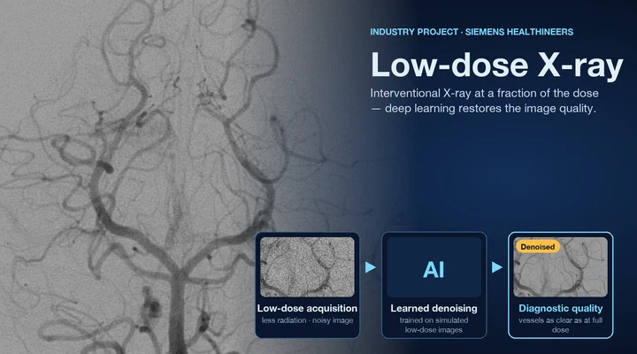
With Siemens Healthineers we reduced the X-ray dose of interventional imaging without losing image quality: photon recycling and noise-model-driven denoising of ultra-low-dose fluoroscopy, low-rank denoising of digital subtraction angiography, and a real-time network trained on simulated low-dose images. Earlier work revealed hidden structures in single X-ray images through X-ray in-depth decomposition and single-view depth recovery for image-guided interventions.
Devices, catheters and stents
Knowing where devices are is essential during an intervention. We detected catheter electrodes and predicted their depth from a single view (CathNets), and segmented stents and multiple devices in fluoroscopy for endovascular aortic repair, and we co-edited the proceedings of the MICCAI workshops on intravascular imaging and computer-assisted stenting (CVII-STENT).
Endoscopic and surgical video
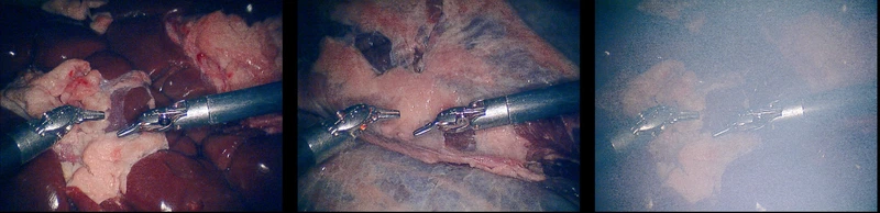
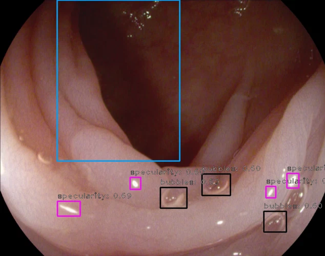
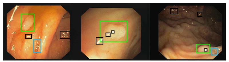
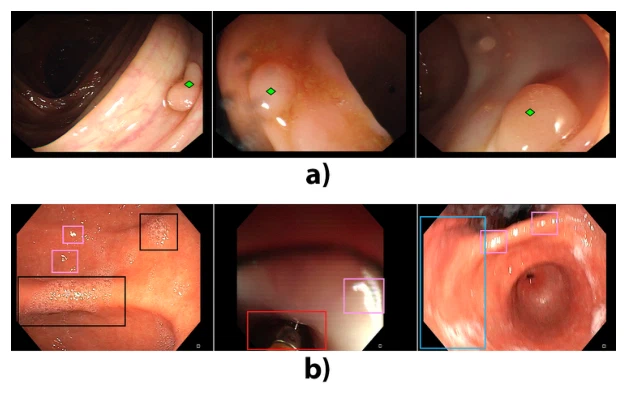
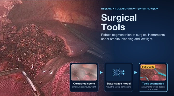
In endoscopy we took part in the Endoscopy Artefact Detection challenge and studied how artefacts such as blur, bubbles and specular reflections affect automated polyp detection — and how to make detectors robust to them. In surgical video, the SegSTRONG-C challenge (IEEE TMI 2026) benchmarked robust surgical tool segmentation under realistic corruptions such as smoke, bleeding and blur, and with Kyung Hee University our surgical tools project explored state-space vision models for robust instrument segmentation.
The common thread is robustness: models that keep working under the noise, artefacts and corruptions that are normal in the interventional suite and the operating room.
 completed