MRI
MRIQT — Image-Quality Transfer for Ultra-Low-Field MRI
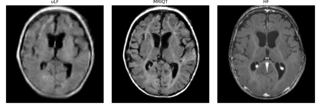
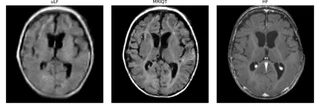
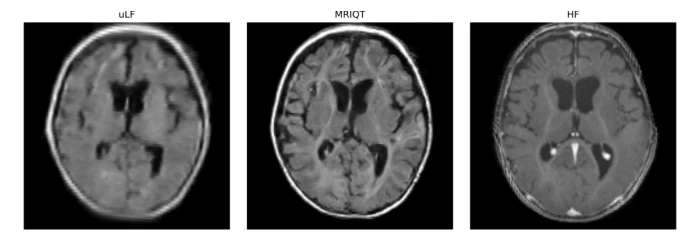
Slide between a portable ultra-low-field (0.064 T) neonatal MRI scan and its MRIQT enhancement — a physics-aware 3D diffusion model that transfers high-field image quality to bedside scans.
Partners




Research themes
Projects


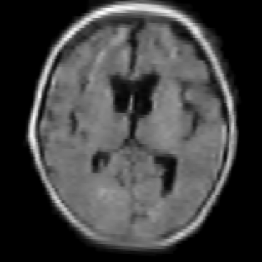
Slide to compare the portable ultra-low-field scan (uLF, right) with the MRIQT enhancement (left). Use the buttons to switch between two patients.
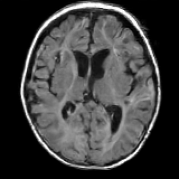 MRIQT
uLF (0.064 T)
arXiv · PDF · Code · Accepted at IEEE ISBI 2026, London
Why it matters
Infants change fast in the first weeks of life: their brains differ from adult brains in both anatomy and tissue contrast. In neonatal intensive care units, many infants have neurological conditions that need repeated MRI, yet standard high-field MRI means disconnecting them from life support and moving them to a scanner room — slow, often requiring sedation and exposure to loud noise.
Portable ultra-low-field MRI (0.064 T) brings the scanner to the bedside, but with low signal-to-noise and reduced diagnostic quality. MRIQT closes that gap: it performs 3D image-quality transfer (uLF → high-field) with a conditional diffusion model guided by physics-aware k-space simulation and an anatomy-preserving 3D perceptual objective. The cohort comprises neonates with an average age of two months and nine clinically relevant neurological pathologies, often occurring together.
How it works
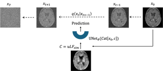
- Physics-aware degradation: realistic ultra-low-field scans are simulated from high-field ones in k-space.
- 3D conditional diffusion: the model enhances the scan while preserving anatomy.
- Guidance and an SNR-weighted 3D perceptual loss reduce artefacts and hallucinations.
Training — MRIQT learns from physics-based uLF simulations to reconstruct anatomically faithful images from noisy inputs:
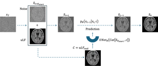
Inference — given a real uLF scan, MRIQT iteratively denoises a noisy initialisation while conditioning on the uLF measurement:
Results
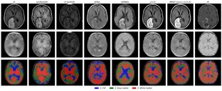
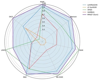
Compared with competing methods on PSNR, LPIPS, MAE, MS-SSIM, SSIM and Pearson correlation:
Related publications
MRIQT: Physics-Aware Diffusion Model for Image Quality Transfer in Neonatal Ultra-Low-Field MRI
2026 IEEE 23rd International Symposium on Biomedical Imaging (ISBI), 2025
How to cite
@inproceedings{alabed2026mriqt,
  title     = {MRIQT: Physics-Aware Diffusion Model for Image Quality Transfer in Neonatal Ultra-Low-Field MRI},
  author    = {Al Abed, Malek and Demir, Sebiha and Groteklaes, Anne and Germani, Elodie and Faghihroohi, Shahrooz and Sabir, Hemmen and Albarqouni, Shadi},
  booktitle = {IEEE International Symposium on Biomedical Imaging (ISBI)},
  year      = {2026},
  eprint    = {2511.13232},
  archivePrefix = {arXiv}
}