Conference
CORE A
❝ citations
X-ray in-depth decomposition: Revealing the latent structures
Medical Image Computing and Computer Assisted Intervention – MICCAI 2017, Lecture Notes in Computer Science, 2017
Abstract
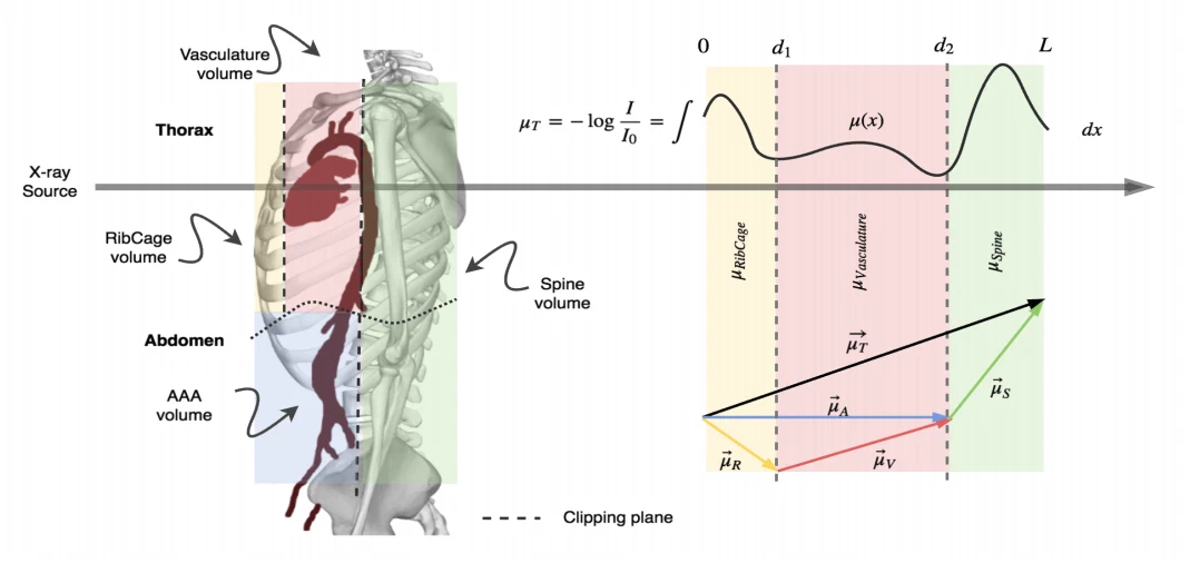
X-ray is the most readily available imaging modality and has a broad range of applications that spans from diagnosis to intra-operative guidance in cardiac, orthopedics, and trauma procedures. Proper interpretation of the hidden and obscured anatomy in X-ray images remains a challenge and often requires high radiation dose and imaging from several perspectives. In this work, we aim at decomposing the conventional X-ray image into d X-ray components of independent, non-overlapped, clipped sub-volume, that separate rigid structures into distinct layers, leaving all deformable organs in one layer, such that the sum resembles the original input. Our proposed model is validaed on 6 clinical datasets (∼7200 X-ray images) in addition to 615 real chest X-ray images. Despite the challenging aspects of modeling such a highly ill-posed problem, exciting and encouraging results are obtained paving the path for further contributions in this direction.
Interactive explainer
Coming soon. An interactive Slidev walkthrough of this paper — with live, explorable figures — will appear here.
Authors: set
slidev: in this page's front matter to the deployed deck URL.Research themes
Keywords
RadiologyMedical ImagingDeep Learning
Lab authors


Cite (BibTeX)
@inproceedings{albarqouni2017x,
 author = {Albarqouni, Shadi and Fotouhi, Javad and Navab, Nassir},
 booktitle = {International Conference on Medical Image Computing and Computer-Assisted Intervention},
 organization = {Springer, Cham},
 pages = {444--452},
 title = {X-ray in-depth decomposition: Revealing the latent structures},
 year = {2017}
}
Related publications
Semi-supervised deep learning for fully convolutional networks
Medical Image Computing and Computer Assisted Intervention – MICCAI 2017, Lecture Notes in Computer Science, 2017
Cathnets: detection and single-view depth prediction of catheter electrodes
Medical Imaging and Augmented Reality (MIAR 2016), Lecture Notes in Computer Science, 2016
X-Ray PoseNet: 6 DoF pose estimation for mobile X-Ray devices
2017 IEEE Winter Conference on Applications of Computer Vision (WACV), 2017