Workshop
❝ 92 citations
Generating highly realistic images of skin lesions with GANs
MICCAI 2018 Workshops (ISIC Skin Image Analysis), LNCS 11041, 2018
Abstract
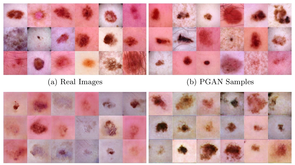
As many other machine learning driven medical image analysis tasks, skin image analysis suffers from a chronic lack of labeled data and skewed class distributions, which poses problems for the training of robust and well-generalizing models. The ability to synthesize realistic looking images of skin lesions could act as a reliever for the aforementioned problems. Generative Adversarial Networks (GANs) have been successfully used to synthesize realistically looking medical images, however limited to low resolution, whereas machine learning models for challenging tasks such as skin lesion segmentation or classification benefit from much higher resolution data. In this work, we successfully synthesize realistically looking images of skin lesions with GANs at such high resolution. Therefore, we utilize the concept of progressive growing, which we both quantitatively and qualitatively compare to other GAN architectures such as the DCGAN and the LAPGAN. Our results show that with the help of progressive growing, we can synthesize highly realistic dermoscopic images of skin lesions that even expert dermatologists find hard to distinguish from real ones.
Interactive explainer
Auto-generated. This explainer was generated automatically with Claude (Anthropic) from the published paper. It simplifies the method and may contain errors — for a correct understanding, and for all results and numbers, please refer to the published paper (publisher version).
Research themes
Keywords
Medical ImagingDeep LearningRadiologyGenerative Adversarial NetworksAnomaly Detection
Lab authors


Cite (BibTeX)
@incollection{baur2018generating,
 author = {Baur, Christoph and Albarqouni, Shadi and Navab, Nassir},
 booktitle = {OR 2.0 Context-Aware Operating Theaters, Computer Assisted Robotic Endoscopy, Clinical Image-Based Procedures, and Skin Image Analysis},
 pages = {260--267},
 publisher = {Springer, Cham},
 title = {Generating highly realistic images of skin lesions with GANs},
 year = {2018}
}
Related publications
Deep autoencoding models for unsupervised anomaly segmentation in brain MR images
Brainlesion: Glioma, Multiple Sclerosis, Stroke and Traumatic Brain Injuries — BrainLes 2018 workshop @ MICCAI, Lecture Notes in Computer Science, 2018

Intraoperative stent segmentation in X-ray fluoroscopy for endovascular aortic repair
International journal of computer assisted radiology and surgery, 2018

Multiple device segmentation for fluoroscopic imaging using multi-task learning
MICCAI 2018 Workshops (CVII-STENT and LABELS), LNCS 11043, 2018
Weakly-supervised localization and classification of proximal femur fractures
arXiv preprint arXiv:1809.10692, 2018