Journal
❝ citations
Diagnostic assessment of deep learning algorithms for detection of lymph node metastases in women with breast cancer
Jama, 2017
Abstract
Importance: Application of deep learning algorithms to whole-slide pathology images can potentially improve diagnostic accuracy and efficiency. Objective: Assess the performance of automated deep learning algorithms at detecting metastases in hematoxylin and eosin-stained tissue sections of lymph nodes of women with breast cancer and compare it with pathologists’ diagnoses in a diagnostic setting. Design, Setting, and Participants: Researcher challenge competition (CAMELYON16) to develop automated solutions for detecting lymph node metastases (November 2015-November 2016). A training data set of whole-slide images from 2 centers in the Netherlands with (n = 110) and without (n = 160) nodal metastases verified by immunohistochemical staining were provided to challenge participants to build algorithms. Algorithm performance was evaluated in an independent test set of 129 whole-slide images (49 with and 80 without metastases). The same test set of corresponding glass slides was also evaluated by a panel of 11 pathologists with time constraint (WTC) from the Netherlands to ascertain likelihood of nodal metastases for each slide in a flexible 2-hour session, simulating routine pathology workflow, and by 1 pathologist without time constraint (WOTC). Exposures: Deep learning algorithms submitted as part of a challenge competition or pathologist interpretation. Main Outcomes and Measures: The presence of specific metastatic foci and the absence vs presence of lymph node metastasis in a slide or image using receiver operating characteristic curve analysis. The 11 pathologists participating in the simulation exercise rated their diagnostic confidence as definitely normal, probably normal, equivocal, probably tumor, or definitely tumor. Results: The area under the receiver operating characteristic curve (AUC) for the algorithms ranged from 0.556 to 0.994. The top-performing algorithm achieved a lesion-level, true-positive fraction comparable with that of the pathologist WOTC (72.4% [95% CI, 64.3%-80.4%]) at a mean of 0.0125 false-positives per normal whole-slide image. For the whole-slide image classification task, the best algorithm (AUC, 0.994 [95% CI, 0.983-0.999]) performed significantly better than the pathologists WTC in a diagnostic simulation (mean AUC, 0.810 [range, 0.738-0.884]; P < .001). The top 5 algorithms had a mean AUC that was comparable with the pathologist interpreting the slides in the absence of time constraints (mean AUC, 0.960 [range, 0.923-0.994] for the top 5 algorithms vs 0.966 [95% CI, 0.927-0.998] for the pathologist WOTC). Conclusions and Relevance: In the setting of a challenge competition, some deep learning algorithms achieved better diagnostic performance than a panel of 11 pathologists participating in a simulation exercise designed to mimic routine pathology workflow; algorithm performance was comparable with an expert pathologist interpreting whole-slide images without time constraints. Whether this approach has clinical utility will require evaluation in a clinical setting.
Interactive explainer
Coming soon. An interactive Slidev walkthrough of this paper — with live, explorable figures — will appear here.
Authors: set
slidev: in this page's front matter to the deployed deck URL.Research themes
Keywords
Deep LearningMedical ImagingHistology
Lab authors


Cite (BibTeX)
@article{bejnordi2017diagnostic,
 author = {Bejnordi, Babak Ehteshami and Veta, Mitko and Van Diest, Paul Johannes and Van Ginneken, Bram and Karssemeijer, Nico and Litjens, Geert and Van Der Laak, Jeroen AWM and Hermsen, Meyke and Manson, Quirine F and Balkenhol, Maschenka and others},
 journal = {Jama},
 number = {22},
 pages = {2199--2210},
 publisher = {American Medical Association},
 title = {Diagnostic assessment of deep learning algorithms for detection of lymph node metastases in women with breast cancer},
 volume = {318},
 year = {2017}
}
Related publications
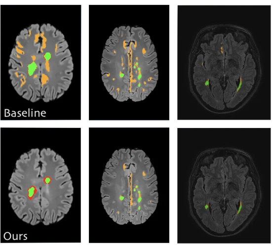
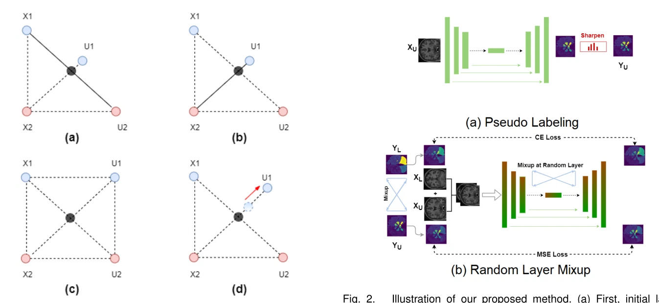
Semi-supervised deep learning for fully convolutional networks
Medical Image Computing and Computer Assisted Intervention – MICCAI 2017, Lecture Notes in Computer Science, 2017
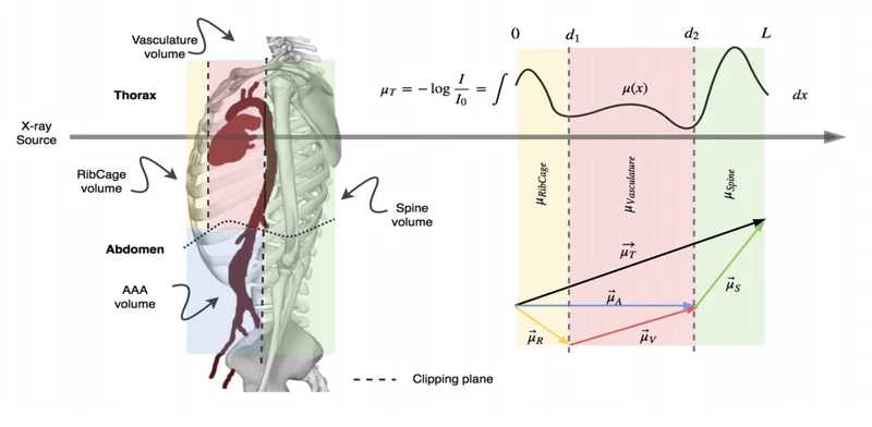
X-ray in-depth decomposition: Revealing the latent structures
Medical Image Computing and Computer Assisted Intervention – MICCAI 2017, Lecture Notes in Computer Science, 2017
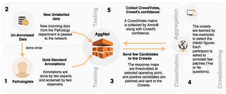
Aggnet: deep learning from crowds for mitosis detection in breast cancer histology images
IEEE transactions on medical imaging, 2016
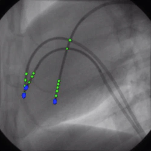
Cathnets: detection and single-view depth prediction of catheter electrodes
Medical Imaging and Augmented Reality (MIAR 2016), Lecture Notes in Computer Science, 2016