Liver lesion localisation and classification with convolutional neural networks: a comparison between conventional and spectral computed tomography
Biomedical Physics & Engineering Express, 2020
Altmetric · top 25% of all research outputs · 2 patents
Abstract
Abstract
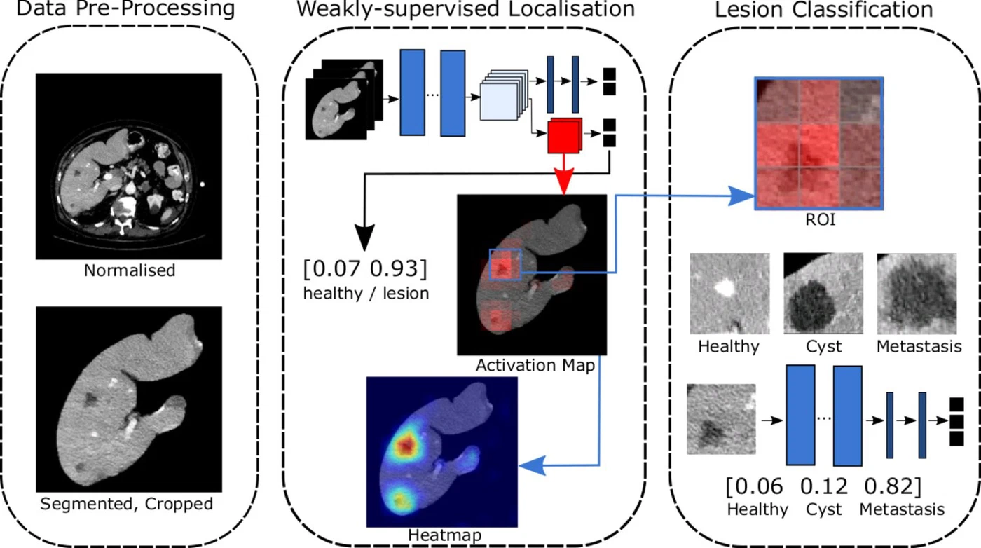
Purpose: To evaluate the benefit of the additional available information present in spectral CT datasets, as compared to conventional CT datasets, when utilizing convolutional neural networks for fully automatic localisation and classification of liver lesions in CT images. Materials and Methods: Conventional and spectral CT images (iodine maps, virtual monochromatic images (VMI)) were obtained from a spectral dual-layer CT system. Patient diagnosis were known from the clinical reports and classified into healthy, cyst and hypodense metastasis. In order to compare the value of spectral versus conventional datasets when being passed as input to machine learning algorithms, we implemented a weakly-supervised convolutional neural network (CNN) that learns liver lesion localisation without pixel-level ground truth annotations. Regions-of-interest are selected automatically based on the localisation results and are used to train a second CNN for liver lesion classification (healthy, cyst, hypodense metastasis). The accuracy of lesion localisation was evaluated using the Euclidian distances between the ground truth centres of mass and the predicted centres of mass. Lesion classification was evaluated by precision, recall, accuracy and F1-Score. Results: Lesion localisation showed the best results for spectral information with distances of 8.22 ± 10.72 mm, 8.78 ± 15.21 mm and 8.29 ± 12.97 mm for iodine maps, 40 keV and 70 keV VMIs, respectively. With conventional data distances of 10.58 ± 17.65 mm were measured. For lesion classification, the 40 keV VMIs achieved the highest overall accuracy of 0.899 compared to 0.854 for conventional data. Conclusion: An enhanced localisation and classification is reported for spectral CT data, which demonstrates that combining machine-learning technology with spectral CT information may in the future improve the clinical workflow as well as the diagnostic accuracy.
Interactive explainer
slidev: in this page's front matter to the deployed deck URL.In short. Spectral (dual-layer) CT gives radiologists more than a single conventional CT image: from one scan it can produce iodine maps and virtual monochromatic images at different energies. We asked whether this extra information also helps deep learning. On abdominal CT scans of patients who were healthy, had liver cysts or had hypodense metastases, we trained the same two-stage pipeline once on conventional images and once on each spectral reconstruction.
How it works. A first convolutional network learns where lesions are without any pixel-level annotations: it is trained only on whether a slice contains a lesion (weak supervision), and its activation maps point to the suspicious regions. Patches around these regions are then passed to a second network that classifies them as healthy tissue, cyst or metastasis.
What we found. Spectral data helped at both steps. Lesions were localised more precisely on iodine maps and low-energy (40 keV) virtual monochromatic images, about 8 mm from the true lesion centre on average, than on conventional images (about 11 mm). For classification, the 40 keV images reached an accuracy of 0.90, against 0.85 for conventional CT. Combining machine learning with spectral CT could therefore make liver lesion assessment both faster and more accurate in clinical practice.
The work was a collaboration between the Department of Diagnostic and Interventional Radiology at Klinikum rechts der Isar (TU Munich), led by Peter B. Noël, and the Chair for Computer Aided Medical Procedures (CAMP) at TU Munich. The article is open access in Biomedical Physics & Engineering Express 6(1), 015038, published 31 January 2020.
Research themes
Research areas
Keywords
Lab authors


Cite (BibTeX)
@article{shapira2020liver,
 author = {Shapira, Nadav and Fokuhl, Julia and Schultheiß, Manuel and Beck, Stefanie and Kopp, Felix K and Pfeiffer, Daniela and Dangelmaier, Julia and Pahn, Gregor and Sauter, Andreas P and Renger, Bernhard and others},
 journal = {Biomedical Physics & Engineering Express},
 number = {1},
 pages = {015038},
 publisher = {IOP Publishing},
 title = {Liver lesion localisation and classification with convolutional neural networks: a comparison between conventional and spectral computed tomography},
 volume = {6},
 year = {2020}
}
Related publications

Benefit of dual energy CT for lesion localization and classification with convolutional neural networks
Medical Imaging 2020: Physics of Medical Imaging, 2020
Towards an Interactive and Interpretable CAD System to Support Proximal Femur Fracture Classification
arXiv preprint arXiv:1902.01338, 2019
Weakly-supervised localization and classification of proximal femur fractures
arXiv preprint arXiv:1809.10692, 2018
Image-to-Images Translation for Multi-Task Organ Segmentation and Bone Suppression in Chest X-Ray Radiography
IEEE Transactions on Medical Imaging, 2020