Journal
Q1IF 12.4
❝ citations
Structure-preserving color normalization and sparse stain separation for histological images
IEEE transactions on medical imaging, 2016
Abstract
Staining and scanning of tissue samples for microscopic examination is fraught with undesirable color variations arising from differences in raw materials and manufacturing techniques of stain vendors, staining protocols of labs, and color responses of digital scanners. When comparing tissue samples, color normalization and stain separation of the tissue images can be helpful for both pathologists and software. Techniques that are used for natural images fail to utilize structural properties of stained tissue samples and produce undesirable color distortions. The stain concentration cannot be negative. Tissue samples are stained with only a few stains and most tissue regions are characterized by at most one effective stain. We model these physical phenomena that define the tissue structure by first decomposing images in an unsupervised manner into stain density maps that are sparse and non-negative. For a given image, we combine its stain density maps with stain color basis of a pathologist-preferred target image, thus altering only its color while preserving its structure described by the maps. Stain density correlation with ground truth and preference by pathologists were higher for images normalized using our method when compared to other alternatives. We also propose a computationally faster extension of this technique for large whole-slide images that selects an appropriate patch sample instead of using the entire image to compute the stain color basis.
Interactive explainer
Coming soon. An interactive Slidev walkthrough of this paper — with live, explorable figures — will appear here.
Authors: set
slidev: in this page's front matter to the deployed deck URL.Research themes
Keywords
HistologyMedical Imaging
Lab authors


Cite (BibTeX)
@article{vahadane2016structure,
 author = {Vahadane, Abhishek and Peng, Tingying and Sethi, Amit and Albarqouni, Shadi and Wang, Lichao and Baust, Maximilian and Steiger, Katja and Schlitter, Anna Melissa and Esposito, Irene and Navab, Nassir},
 journal = {IEEE transactions on medical imaging},
 number = {8},
 pages = {1962--1971},
 publisher = {IEEE},
 title = {Structure-preserving color normalization and sparse stain separation for histological images},
 volume = {35},
 year = {2016}
}
Related publications
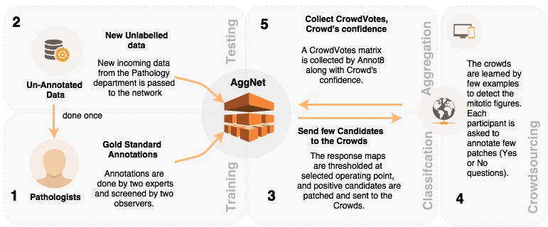
Aggnet: deep learning from crowds for mitosis detection in breast cancer histology images
IEEE transactions on medical imaging, 2016

Structure-preserved color normalization for histological images
2015 IEEE 12th International Symposium on Biomedical Imaging (ISBI), 2015
Steps towards Establishing Telemedicine Center in Palestine
2nd International Conference on e-Health and Bioengineering (EHB 2009), Iași–Constanța, Romania, 2009
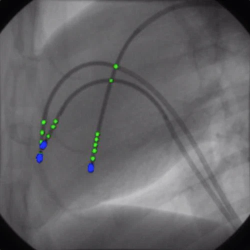
Cathnets: detection and single-view depth prediction of catheter electrodes
Medical Imaging and Augmented Reality (MIAR 2016), Lecture Notes in Computer Science, 2016