Benefit of dual energy CT for lesion localization and classification with convolutional neural networks

Abstract
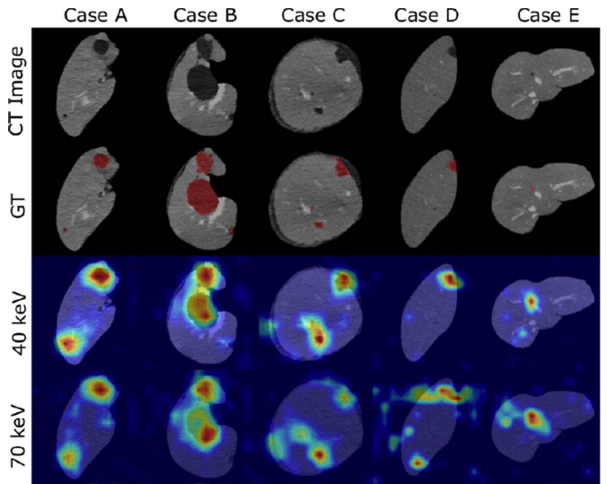
Dual Energy CT is a modern imaging technique that is utilized in clinical practice to acquire spectral information for various diagnostic purposes including the identification, classification, and characterization of different liver lesions. It provides additional information that, when compared to the information available from conventional CT datasets, has the potential to benefit existing computer vision techniques by improving their accuracy and reliability. In order to evaluate the additional value of spectral versus conventional datasets when being used as input for machine learning algorithms, we implemented a weakly-supervised Convolutional Neural Network (CNN) that learns liver lesion localization and classification without pixel-level ground truth annotations. We evaluated the lesion classification (healthy, cyst, hypodense metastasis) and localization performance of the network for various conventional and spectral input datasets obtained from the same CT scan. The best results for lesion localization were found for the spectral datasets with distances of 8.22 ± 10.72 mm, 8.78 ± 15.21 mm and 8.29 ± 12.97 mm for iodine maps, 40 keV and 70 keV virtual mono-energetic images, respectively, while lesion localization distances of 10.58 ± 17.65 mm were measured for the conventional dataset. In addition, the 40 keV virtual mono-energetic datasets achieved the highest overall lesion classification accuracy of 0.899 compared to 0.854 measured for the conventional datasets. The enhanced localization and classification results that we observed for spectral CT data demonstrates that combining machine-learning technology with spectral CT information may improve the clinical workflow as well as the diagnostic accuracy.