Unsupervised Deep Learning for Medical Image Analysis is increasingly gaining attention, since it relieves from the need for annotating training data. Recently, deep generative …
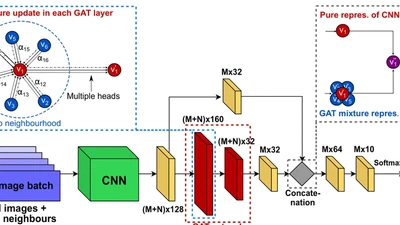
Recently, Geometric Deep Learning (GDL) has been introduced as a novel and versatile framework for computer-aided disease classification. GDL uses patient meta-information such as …
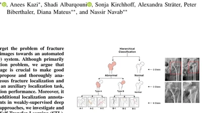
In this paper, we target the problem of fracture classification from clinical X-Ray images towards an automated Computer Aided Diagnosis (CAD) system. Although primarily dealing …
For endovascular aortic repair (EVAR), integrating preoperative information of the aortic anatomy with intraoperative fluoroscopy can aid in reducing radiation exposure, contrast …
Fusion of preoperative data with intraoperative X-ray images has proven the potential to reduce radiation exposure and contrast agent, especially for complex endovascular aortic …
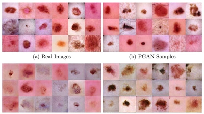
As many other machine learning driven medical image analysis tasks, skin image analysis suffers from a chronic lack of labeled data and skewed class distributions, which poses …
Segmentation of the left atrium and deriving its size can help to predict and detect various cardiovascular conditions. Automation of this process in 3D Ultrasound image data is …
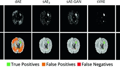
Reliably modeling normality and differentiating abnormal appearances from normal cases is a very appealing approach for detecting pathologies in medical images. A plethora of such …
Clinical procedures that make use of fluoroscopy may expose patients as well as the clinical staff (throughout their career) to non-negligible doses of radiation. The potential …
Precise reconstruction of 3D volumes from X-ray projections requires precisely pre-calibrated systems where accurate knowledge of the systems geometric parameters is known ahead. …
Privacy — With your consent we use Google Analytics to understand how our website is used (IP addresses anonymised).
You can change your choice at any time via “Cookie settings” in the footer. Privacy policy